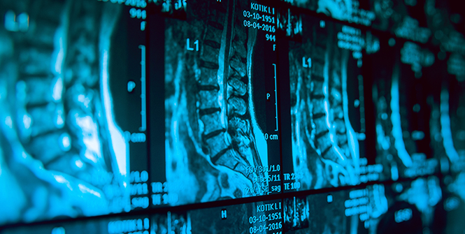
Patients with traumatic spinal cord injury (SCI) often have an unpredictable neurological outcome, and there is a need for more accurate prognostication tools. Imaging assessment of spinal cord damage can potentially predict neurological recovery after acute SCI. Currently, conventional T2-weighted MRI serves as the clinical standard for characterizing injury to the spinal cord parenchyma, which is often a T2-weighted hyperintensity with or without T1-weighted hypointensity at the site of the traumatic lesion. Although conventional MRI provides macrostructural detail sufficient for clinical care and surgical planning, it has limited ability in predicting patient outcome or characterizing neural degeneration along the neuraxis over time. Diffusion MRI of the spinal cord offers the ability to quantify neuronal injury within the spinal cord as well as improve prognostication for SCI.
Diffusion MRI, a magnetic resonance technique which measures diffusion of water molecules in tissues, is sensitive to microstructural changes within the spinal cord at a resolution smaller than the actual image resolution.1 Diffusion tensor imaging (DTI) takes advantage of the preferential diffusion of water molecules along axons and produces metrics such as fractional anisotropy (FA), axial diffusion (AD), radial diffusion (RD), and apparent diffusion coefficient (ADC), which have been associated with axonal degeneration, demyelination, and edema following traumatic SCI in both animal models and humans.2 Importantly, DTI captures microstructural changes in regions of the spinal cord that appear normal on conventional MRI.3 In acute SCI, FA and AD decreases (indicative of axonal injury) at the lesion site, and these metrics correlate with ASIA (American Spinal Injury Association) motor scores.4,5 In chronic SCI, FA is decreased rostral to the lesion site, highlighting the ability of DTI to quantify retrograde Wallerian degeneration in areas of the spinal cord that appear normal on conventional T2-weighted MRI.6 Interestingly, radial diffusivity of the gray matter is increased in the lumbar enlargement in subjects with cervical SCI and this correlates with electrophysiologic and clinical motor scores.7 Together, the use of DTI to assess remote neurodegeneration offers potential applications for diagnosis and prognosis.
DTI enables investigation of pathophysiological changes within the spinal cord after acute SCI. This non-invasive tool can measure microstructural changes in the spinal cord after therapeutic interventions, such as reduction in edema with anti-inflammatory therapies or axonal regeneration following stem cell injection. These applications could lead to the use of DTI metrics as end-point biomarkers in clinical trials. Recent approaches such as filtered diffusion weighted imaging (fDWI) aim to reduce confounders and more accurately measure axonal injury, which is the primary predictor of motor outcomes.8 Additionally, MRI processing software developed specifically for the spinal cord, such as the Spinal Cord Toolbox,9 makes it possible to measure tract-specific neural injury and degeneration.10
Future research in this field is aimed at establishing standardized diffusion MR sequences for spinal cord imaging, which will improve our ability to collect and analyze multi-center data. There is work being done to improve image resolution with spinal cord diffusion MRI as well as reduce metallic artifacts related to spinal instrumentation. In the future, the addition of diffusion MR imaging protocols to standard clinical protocols could offer physicians a tool to monitor regenerative or therapeutic interventions and improve prognostication for acute SCI.
References
- Basser, P. J. & Pierpaoli, C. Microstructural and physiological features of tissues elucidated by quantitative-diffusion-tensor MRI. J. Magn. Reson. B 111, 209–219 (1996).
- Vedantam, A. et al. Diffusion tensor imaging of the spinal cord: insights from animal and human studies. Neurosurgery 74, 1–8; discussion 8; quiz 8 (2014).
- Shen, H. et al. Applications of diffusion-weighted MRI in thoracic spinal cord injury without radiographic abnormality. Int. Orthop. 31, 375–383 (2007).
- Cheran, S. et al. Correlation of MR diffusion tensor imaging parameters with ASIA motor scores in hemorrhagic and nonhemorrhagic acute spinal cord injury. J. Neurotrauma 28, 1881–1892 (2011).
- Shanmuganathan, K., Gullapalli, R. P., Zhuo, J. & Mirvis, S. E. Diffusion tensor MR imaging in cervical spine trauma. AJNR Am. J. Neuroradiol. 29, 655–659 (2008).
- Ellingson, B. M., Ulmer, J. L., Kurpad, S. N. & Schmit, B. D. Diffusion tensor MR imaging in chronic spinal cord injury. AJNR Am J Neuroradiol 29, 1976–1982 (2008).
- David, G. et al. In vivo evidence of remote neural degeneration in the lumbar enlargement after cervical injury. Neurology 92, e1367–e1377 (2019).
- Budde, M. D., Skinner, N. P., Muftuler, L. T., Schmit, B. D. & Kurpad, S. N. Optimizing Filter-Probe Diffusion Weighting in the Rat Spinal Cord for Human Translation. Front. Neurosci. 11, 706 (2017).
- De Leener, B. et al. SCT: Spinal Cord Toolbox, an open-source software for processing spinal cord MRI data. NeuroImage 145, 24–43 (2017).
- David, G. et al. Traumatic and nontraumatic spinal cord injury: pathological insights from neuroimaging. Nat. Rev. Neurol. 15, 718–731 (2019).


