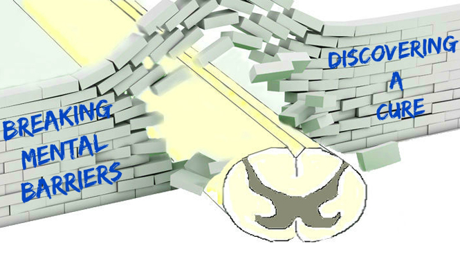
Why don’t we have a cure for spinal cord injury? We can put a man on the moon and obtain encyclopedic knowledge through the air on a handheld device, but we have not been able to cure spinal cord injury. Why is that? One reason may be the incredibly negative attitudes towards trying to repair the spinal cord. Several “doctrines” have been proposed in the past that have significantly hindered advances towards spinal cord injury repair. Fortunately, we have been able to overcome these barriers such that maybe in the future, we will ave a cure for spinal cord injury. We briefly review some of the myths below.
Doctrine: Spinal Cord Injury was an “Ailment not to be Treated”
We know from the Edwin Smith Surgical Papyrus that spinal cord injury was considered an “ailment not to be treated” ever since 2500 to 1900 B.C. (1). Spinal cord injuries were treated in the past with bed rest, which led to numerous complications and a high mortality rate. With the development of improved spinal fixation techniques, nursing care and rehabilitation protocols, we have been able to significantly improve care for patients with spinal cord injury. While we could argue that short-term survival after spinal cord injury has significantly improved since the 1970s (2), the incidence rates of spinal cord injury has remained relatively stable between 1993 and 2012 in the U.S. (3), and we still cannot reliably restore lost function.
Doctrine: Spinal Decompression Alone Will Decompress the Spinal Cord
While we currently cannot restore function, one of the guiding principles of the management of spinal cord injury is decompression of the spinal cord. There has been much study of methods to restore spinal canal diameter, including traction and instrumentation to restore spinal alignment, discectomy, corpectomy and laminectomy. It has been the belief that the restoration of spinal alignment and canal diameter will decompress the spinal cord; however, several studies have found that this may not be enough. Kwon et. al (4) placed lumbar intrathecal catheters into 22 patients with spinal cord injury and surgical decompression. They found that all patients had a cerebrospinal fluid (CSF) waveform pattern that was flat with no pulsations that became pulsatile again post-decompression, suggesting that the surgical decompression did decompress the spinal cord. However, a “number” of patients had their pulsatile waveform revert back to the flap pattern with magnetic resonance imaging (MRI) demonstrating ongoing compression of the cord or spinal cord edema against the dura such that the CSF waveform was no longer transmitted.
Werndle et. al (5) performed intraspinal pressure monitoring of the spinal cord at the injury site after spinal cord injury and found that bony realignment and laminectomy did not effectively lower intraspinal pressure. In fact, laminectomy was detrimental as it exposed the swollen spinal cord to compression forces that are applied to the skin when patients were nursed prone. This group followed up this work by showing that expansion duroplasty improves intraspinal pressure, spinal cord perfusion pressure and vascular pressure reactivity index in patients with traumatic spinal cord injury (6).
Duraplasty to decompress neural structures is intuitive. Neurosurgeons routinely open the dura to decompress injured and edematous brain tissue after injury in selected cases. No neurosurgeon would consider that removal of the skull flap alone, without durotomy, would significantly decompress the brain, and so opening the dura to compress the injured, swollen spinal cord in selected cases would come naturally to neurosurgeons. However, spinal cord-injured patients are also cared for by orthopaedic surgeons with less familiarity with durotomy and duraplasty, and this may impact on future care and cure of patients with spinal cord injury.
Doctrine: Injured Central Nervous System (CNS) Axons Cannot Regenerate
While we have made impact in the care of patients with spinal cord injury, a major impediment has been the “doctrine” that injured CNS axons cannot regenerate. In the early 1900s, Nobel laureate, Santiago Ramon y Cajal (7,8) wrote that the injured CNS axons fail to regenerate, but this was dogma. Research efforts thus have focused on neuroprotection rather than neuroregeneration. The translation of neuroprotection research to humans has resulted in clinical trials that strive to decrease the extent of injury; however, repair is still significantly limited. Indeed, clinical trials involving methylprednisolone (9,10), minocycline (11,12) and riluzole (13,14) are all therapies that involve neuroprotection. While these therapies are laudable for their potential to ameliorate injury, they are a far cry from a cure.
It was not until the 1980s that David, Richardson and Aguayo (15-18) brought hope to the neuroscience community for a cure. Their series of papers demonstrated that the doctrine that spinal cord axons cannot regenerate was incorrect. CNS axons can regenerate provided they are given an environment that does not inhibit regrowth. Much work has been done to identify and neutralize these inhibitors, which has led to some potentially promising clinical trials (see below). Furthermore, the discovery of neural stem cells (19) only increased the fervor that a cure for spinal cord injury and the recovery of function was imminent. Since these reports, there have been a plethora of publications indicating that several cell/tissue transplants (20,21), growth factors (22), biomaterials (23-26) or genetic manipulations( 27,28) or combinations of manipulations (29,30) can result in axonal regeneration and restoration of function. While we still do not have a cure for spinal cord injury, some of these basic science advances are being brought to clinical trials.
Potentially Promising Therapies
While we do not intend to be exhaustive in our list, we have highlighted a few trials to demonstrate some therapies that might contribute to a future curative therapy:
VX-210, Vertex Pharmaceuticals Incorporated
One therapy that is more geared towards neural regeneration as opposed to neuroprotection is VX-210. VX-210 has been shown to inhibit Rho activation, which can then facilitate axon regeneration in the CNS after injury. In vitro and in vivo studies have validated the Rho pathway was an important regulator controlling neuronal response to growth inhibitory proteins after CNS injury. VX-210 was previously marketed as Cethrin and has been assessed in patients. In a preliminary multicenter trial of 48 patients with complete American Spinal Injury Association (ASIA) assessment A injury, VX-210 was given as a single dose during surgery to test the safety and tolerability of the drug, as well as the neurological status of patients following its administration (17). The events that followed were typical of the spinal cord injury population with no serious adverse events attributed to the drug. Both cervical and thoracic spinal cord injury patients were included in the study, but the largest change in motor score was seen in cervical patients treated with 3 mg of VX-210. An improvement of 27.3±13.3 points on the ASIA motor score was observed in this cohort of patients after 12 months of injury. Additionally, 66 percent of patients in this cohort converted from ASIA A to ASIA C or D, suggesting that VX-210 is a promising drug that is able to help patients with neurological recovery after cervical spinal cord injury (17).
Moving forwards, Vertex Pharmaceuticals has begun a phase II/III clinical trial, which has begun recruitment in the U.S. and Canada. Using change from baseline upper extremity motor scores as a primary outcome measure, this clinical trial hopes to recruit 150 patients and is scheduled to complete recruitment by June 2018. More information can be found at clinicaltrials.gov using identifier NCT02669849 (18).
The INSPIRE Study, a Neuro-Spinal Scaffold by InVivo Therapeutics
The Neuro-Spinal Scaffold also targets the regeneration principle of spinal cord repair. Since 84 percent of patients with complete thoracic spinal cord injuries remain at ASIA A for six months post-injury, the Neuro-Spinal Scaffold is an investigational device that is implanted into injured spinal cords to promote tissue remodeling and preservation of spinal cord architecture at the implant site. In the rat model, the Neuro-Spinal Scaffold is shown to preserve spine architecture and has resulted in increased remodeled tissue that is potentially permissive to neural regeneration (19).
In a preliminary study with five patients, a scaffold was implanted between 10 and 83 hours following non-penetrating complete spinal cord injury. No serious adverse effects were shown, and two patients showed significant improvements in AISA classification, one patient showed improvements in sensory zone of preservation and two patients are still awaiting follow-up (20).
The INSPIRE study is continuing recruitment in 20 sites across the U.S. and is projected to be completed around June 2017, with the recruitment of a total of 20 patients. More information on this phase III clinical trial can be found at clinicaltrials.gov using the identifier NCT02138110 (21).
AST-OPC1 by Asteria Biotherapeutics, Inc.
AST-OPC1 administration follows the principle of cell replacement. Laboratory studies have shown that AST-OPC1, which consists of oligodendrocyte progenitor cells produced from human stem cells, promoted partial repair of damaged spinal cord tissue and improved movement of front and back legs in the rat model post spinal cord injury (22). The safety of AST-OPC1 administration was also tested, and no adverse events were seen in the experimental animals, supporting the initiation of a phase I clinical (22).
This clinical trial, dubbed SCI star, is currently in phase I/IIa. Open for recruitment in six U.S. centers, this study is projected to recruit a total of 35 patients and be completed in September 2018. For more information on the phase I/IIb clinical trial, please visit clinicaltrials.gov using identifier NCT02302157 (23).
While there are still some practical barriers that exist, once the mental barriers have been overcome, practical barriers become less insurmountable. We could place a man on the moon because someone believed it could be achieved. We can obtain encyclopedic knowledge through the air on a handheld device because someone believed it could be achieved. While there have been many blockades to the belief that spinal cord injury can be cured, many of these mental obstacles have been overcome, and hopefully, in the near future, we can say that a cure for spinal cord injury has been achieved.
7. Ramón y Cajal, S. Estudios sobre la Degeneración y Regeneración del Sistema Nervioso. (Moya, 1913, 1914).
8. Ramón y Cajal, S. Degeneration and Regeneration of the Nervous System. (Hafner, 1928).
10. Young, W. NASCIS. (1990). National Acute Spinal Cord Injury Study. Journal of neurotrauma, 7, 113-114.
19. Gage, F.H. (2000). Mammalian neural stem cells. Science, 287(5457), 1433-1438.
30. Young, W. (2014) Spinal cord regeneration. Cell transplantation, 23(4-5), 573-611.
31. Clinicaltrials.gov. (April, 2016 The INSPIRE Study: Probable Benefit of the Neuro- Spinal Scaffold for Treatment of AIS A Thoracic Acute Spinal Cord Injury. retrieved from https://clinicaltrials.gov/ct2/show/record/NCT02138110?term=Neuro-spinal+scaffold&rank=1.


