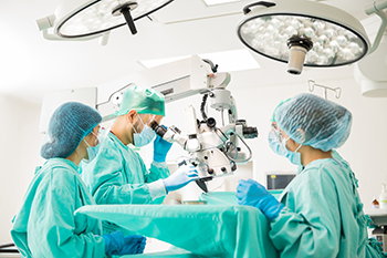
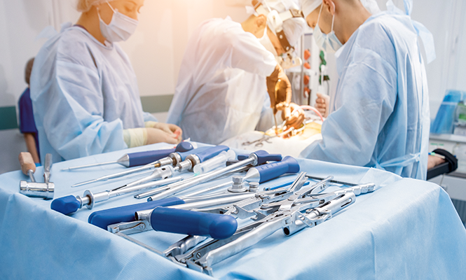
Since the 1800s, even before surgical specialization, surgeons have created new tools to help solve difficult challenges in the operating room. The earliest pioneers in the field of neurosurgery rightfully identified the need for specialized instruments to remove the skull and spinal bones in the most delicate manner possible, giving birth to the wide variety of rongeurs we utilize to this day. With the formal development of the field of neurosurgery, Dr. Harvey Cushing and others contributed to the exponential rise of tools to meet the needs of the specialty. Then Dr. M. Gazi Yasargil ushered in the era of micro-neurosurgery, resulting in another wave of innovation for instrumentation. The familiar call for a Rhoton microinstrument and other eponymous tools can be heard throughout operating rooms, reflecting the rich history of the field and the surgeons of times past and present.
 In early iterations of cranial and spine surgery, the need for efficient, yet precise, removal of bone brought about the invention of rongeurs. “Rongeur” was adapted from a French word, which means “to gnaw”. Dr. Philip D. Kerrison (1872-1944) was an American otologic surgeon who pioneered the Kerrison rongeur (a.k.a. “punch”), originally for decompression of the facial nerve in suppurative otitis media.1,2 This unique instrument was designed with a blunt distal end onto which the sharp end can take a “punch” of bone, therefore removing bone from the inside-out.3 Dr. Lars G. F. Leksell (1907-1986) was a Swedish surgeon well known for his discovery of the gamma motor neuron and invention of Gamma Knife radiosurgery.4 While serving as a battle surgeon in the Finnish Winter War, he developed the Leksell rongeur to quickly and efficiently remove lamina to decompress the spinal cord.5
While rongeurs helped deal with bony decompression, instruments for dissection and elevation were developed to gently manipulate neural tissue. Dr. Wilder G. Penfield (1891-1976), an American-born neurosurgeon, is famously known for his work in neurophysiology and cortical mapping of the motor homunculus still used to this day.6 In the world of neurosurgery, he is famous for his contributions to epilepsy surgery. It was for this work that he developed a set of five blunt-tipped instruments to replace more crude techniques of manipulating tissue with forceps and sponges.7 Similar instruments for blunt manipulation were also borrowed from other specialties, e.g. the Woodson dental instrument (E.W. Woodson) and Freer laryngology elevator (Otto Freer).3,8
As the instruments and the field of neurosurgery expanded, pioneers in spine surgery such as Drs. McCulloch and Cloward brought forth the tools needed to retract muscle and fascia for access of deeper anatomical structures. Dr. John A. McCulloch (1938-2002) was a Canadian orthopedic surgeon who embraced the use of the operating microscope in spinal surgery.9 The steady and unencumbered hands required for this type of surgery necessitated the development of strong, self-retaining retractors; these McCulloch retractors are still used by orthopedic surgeons and neurosurgeons to this day. Dr. Ralph B. Cloward (1908-2000), an American neurosurgeon, was an early pioneer of the anterior cervical discectomy and fusion surgery (ACDF) and developed the handheld Cloward retractors to easily retract the soft tissue of the neck (hand-held) and also the longus colli muscles (self-retaining) for optimizing exposure of the spinal column.3,10
 Starting in the 1950s, when crude operating microscopes were morphing into the feats of engineering they are today, Dr. Yasargil (1925-present) has been a pioneer in microneurosurgery.11,12 With growth in cerebrovascular and neurosurgical oncology, there was a new need for delicate instruments to retract, elevate and dissect, while using the microscope. While many, including Dr. Yasargil himself, have contributed to the development of microinstruments, there is none quite as ubiquitous as the Rhoton micro-dissectors. Dr. Albert L. Rhoton Jr. (1932-2016) was devoted to the study of microneurosurgical anatomy. His contributions, including “Cranial Anatomy and Surgical Approaches” and the Rhoton Collection® (a vast cadaveric anatomic atlas), are still gold-standards in anatomical study.13 The perfection of his well-known microinstruments facilitated in vitro exposure.
From the rongeurs emanating from battlefields to Rhoton micro-dissectors, time and history have required us to develop ingenious new tools to efficiently and delicately accomplish the wide world of neurosurgical procedures. As the field of neurosurgery expands, there will continue to be innovators who continue to enhance the procedures neurosurgeons can safely perform.
References
[expand title=”View All”]
1. Kerrison, P. D. (1904). A bone forceps for use in tympanic surgery; its value in safe-guarding the facial nerve in the radical operation for chronic suppurative otitis media. The Laryngoscope, 14(5), 337–345. doi: 10.1288/00005537-190405000-00001
2. American Otological Society. (1944). In Memoriam: Philip D. Kerrison (1861-1944). AOS Transactions, 33, 321. Retrieved from https://www.dropbox.com/s/ky0lm1rqjplec8a/1944.pdf?dl=0
3. Buraimoh, M., Basheer, A., Taliaferro, K., Shaw, J. H., Haider, S., Graziano, G., & Koh, E. (2018). Origins of eponymous instruments in spine surgery. Journal of Neurosurgery: Spine, 29(6), 696–703. doi: 10.3171/2018.5.spine17981
4. Leksell L. (1949). A stereotaxic apparatus for intracerebral surgery. Acta Chirurgica Scandinavica. 99(3), 229-233.
5. Lindquist, C., & Kihlström, L. (1996). Department of Neurosurgery, Karolinska Institute: 60 Years. Neurosurgery, 39(5), 1016–1021. doi: 10.1227/00006123-199611000-00026
6. Todman, D. (2008). Wilder Penfield (1891–1976). Journal of Neurology, 255(7), 1104–1105. doi: 10.1007/s00415-008-0915-6
7. Penfield, W. (1977). No man alone: a neurosurgeons life. Boston: Little, Brown.
8. Chittiboina, P., Connor, D. E., & Nanda, A. (2012). Dr. Otto “Tiger” Freer: inventor and innovator. Neurosurgical Focus, 33(2). doi: 10.3171/2012.6.focus12137
9. Fraser, R. (2002). In Memoriam: John McCulloch, MD, FRCSC (1938-2002). Spine, 27(21), 2418. doi: 10.1097/00007632-200211010-00024
10. Maiti, T. K., Konar, S. K., Bir, S. C., Kalakoti, P., & Nanda, A. (2016). Ralph Bingham Cloward (1908–2000): Spine Polymath. World Neurosurgery, 89, 562–567. doi: 10.1016/j.wneu.2015.06.061
11. Ya?argil, M. G. (2010). Personal considerations on the history of microneurosurgery. Journal of Neurosurgery, 112(6), 1163–1175. doi: 10.3171/2009.7.jns091124
12. Rogers, L. (2015). Gazi Yasargil: father of modern neurosurgery. Virginia Beach, VA: Ko?ehlerbooks.
13. Matsushima, T., Matsushima, K., Kobayashi, S., Lister, J. R., & Morcos, J. J. (2018). The microneurosurgical anatomy legacy of Albert L. Rhoton Jr., MD: an analysis of transition and evolution over 50 years. Journal of Neurosurgery, 129(5), 1331–1341. doi: 10.3171/2017.7.jns17517
[/expand]
[aans_authors]


