Simple Decompression Versus Anterior Transposition for Cubital Tunnel Syndrome
CASE DESCRIPTION
This 58-year-old, right-handed, hypertensive but otherwise healthy gentleman presented with right hand fourth and fifth finger numbness and tingling, weakness of right hand grip strength and difficulty with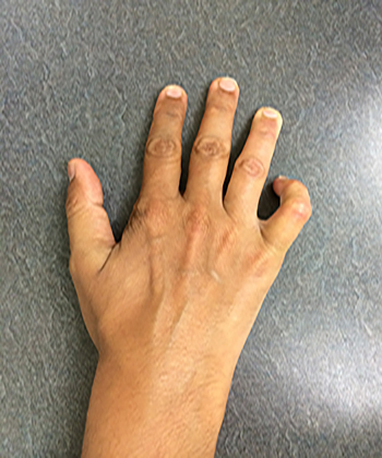 fine hand movements, such as buttoning his shirt. He reported his fifth finger getting caught in the seam of his pant pocket when he tried to put his hand there. He denied any neck or arm pain and had no right upper extremity symptoms. His symptoms, progressively worsening over the past 12 months, had not improved despite use of non-steroidal, anti-inflammatory medications (NSAIDs).

On physical exam of the right hand, 3/5 weakness was noted in the adductor pollicis and abductor digiti minimi, along with wasting and atrophy of the hypothenar and first dorsal interosseous muscle groups. At rest, mild clawing of the 4th and 5th fingers was noted. He was unable to pinch a piece of paper between his first finger and thumb without flexing the distal phalanxes (Positive Froment’s sign) and had percussion tenderness and paresthesias in the ulnar nerve distribution (Positive Tinel’s sign at the cubital tunnel). Loss of sensation in the ulnar distribution of the hand, including the medial half of the fourth finger, the fifth finger and the volar aspect of the palm, was noted. No signs suggestive of Horner’s syndrome were noted, and deep tendon reflexes were normal without clinical signs of radiculopathy. There were no fasciculations of the tongue.
EMG/NCV studies demonstrated slowing of conduction in the right ulnar nerve at the cubital tunnel with low amplitude dorsal ulnar sensory nerve action potential (SNAP) and compound muscle action potentials (CMAP) recording at the first dorsal interrosseus and hypothenar muscles. Fibrillations in ulnar nerve supplied muscles and decreased motor unit action potentials (MUAP) in the hypothenar muscles and the adductor pollicis.
[aans_poll id=”4914″ view=”prompt”]
Discussion
Compromise of the ulnar nerve at the elbow in the retrocondylar groove and cubital tunnel is the second most common compressive neuropathy. Entrapment of the nerve occurs primarily as it traverses the cubital tunnel between the medial epicondyle of the humerus and the olecranon process of the ulna. The roof of this tunnel overlying the ulnar nerve is the thick arcuate fascia and ligament between these two structures. Along with the fascia between the two heads of the flexor carpi ulnaris (FCU), this constitutes Osbourne’s ligament. Additional fibrous bands in this region may also contribute to compression of the ulnar nerve in the cubital tunnel. In addition, the process is dynamic with increased tautness of Osbourne’s ligament when the elbow is flexed contributing to increased ulnar nerve compression in this position. Conversely, extension of the elbow relaxes the fascial bands and ligament relieving compression on the nerve.
The anconeus epitrochlearis, an anomalous muscle found in 10 percent of individuals, spans the interval between the medial epicondyle and olecranon process and may also contribute to ulnar nerve compression in this region; once again, increased compression is noted when the elbow is flexed. Hypertrophy of this muscle may also be seen at times, particularly in athletes, and that may be the sole constricting agent. The Arcade of Struthers is a thin aponeurotic band approximately 8 cm proximal to the elbow that extends between the medial head of the triceps and the medial intermuscular septum. In most instances, the ulnar nerve passes below this as it pierces the medial intermuscular septum and descends over the medial head of the triceps to enter the cubital tunnel. Although purported to cause ulnar nerve compression, this is rarely the case when the nerve is in its native position; in fact, Struther’s original description of these mid-arm compressive fascial bands did not mention this arcade or its role in ulnar nerve compression.
Patients with cubital tunnel syndrome generally present with paresthesias along the medial half of the fourth finger, the fifth finger and the hypothenar eminence and adjacent aspect of the palm. This betrays involvement of the nerve proximal to the origin of the palmar cutaneous branch of the ulnar nerve; this would not be the case with compression of the nerve at Guyon’s canal. Weakness of hypothenar muscles and the adductor pollicis is characteristic, and muscle atrophy in notable in more severe cases with a “claw” deformity of the fourth and fifth fingers at rest. Weakness and wasting of the first dorsal interosseous muscle is noted in advanced cases. Pain is usually not a presenting symptom, but patients may report elbow discomfort and the “funny bone” painful paresthesias. Wartenberg’s sign, ulnar deviation of the fifth finger (weakness of the palmar interossei cause inability to completely adduct the small finger) and Froment’s sign (failure to grip a piece of paper due to weakness of the adductor pollicis) may be seen.
The clinical presentation and electrophysiological studies bring forth the definitive diagnosis of cubital tunnel syndrome with compromise of the ulnar nerve in this location, as seen in this case. The treatment for mild to moderate cubital tunnel syndrome may consist of conservative measures such as simple activity modification, splinting to reduce repetitive elbow flexion and nerve glide techniques often performed by physical therapists. A failure to improve or further decline in ulnar nerve function, particularly the onset of muscle weakness and wasting, prompts a consideration for surgical intervention.
The definitive treatment for persistently symptomatic cubital tunnel syndrome is surgery. There are a number of surgical techniques that have been developed and utilized, but two of the most common are simple decompression of the cubital tunnel and decompression combined with anterior transposition of the ulnar nerve. There is a fair amount of debate and uncertainty regarding which surgical technique yields the best results and patient outcomes. A number of observational studies and randomized control trials comparing surgical approaches for cubital tunnel syndrome have been performed over the last 10 years.
In 2005, Nabhan et al. compared outcomes of 66 patients with cubital tunnel syndrome prospectively randomized to undergo a simple decompression (32 patients) or subcutaneous anterior transposition (34 patients). Follow up at three and nine months showed that both groups of patients experienced significant improvement in symptoms; decreased pain, reduced sensory symptoms and improvement in muscle strength. There was also clearly demonstrated improvement in ulnar nerve electrophysiological studies in both groups. However, there was no statistically significant difference in the degree of symptom improvement and outcomes between the two surgical cohorts.
Also in 2005, Gervasio et al. compared outcomes in 70 patients with severe cubital tunnel syndrome who underwent simple decompression (35 patients) versus submuscular anterior transposition (35 patients). Subjective parameters such as functional outcome and return to work status, as well as improvement in symptoms and objective measures such as grip strength and performance on sensory tests were assessed. Both patient groups experienced significant improvement in their symptoms. Of the patients who underwent simple decompression, 54 percent rated their outcome as excellent, 26 percent as good and 20 percent as fair. Outcomes of patients undergoing submuscular anterior transposition were rated as 51 percent excellent, 31 percent as good and 17 percent as fair. All patients experienced significant improvement in the symptoms immediately following surgery. There was no statistically significant difference in outcome between the two groups, and they noted equivalent efficacy between the two surgical approaches. Electrophysiological outcomes also demonstrated a statistically significant improvement in both groups; again, there was no statistical difference between patients who underwent decompression versus transposition. The results of this study suggest that both simple decompression and submuscular anterior transposition are equally effective methods for treating severe cubital tunnel syndrome.
In 2008, Macadam et al. performed a meta-analysis of 10 randomized controlled trials and comparative observational studies comparing simple decompression to either subcutaneous anterior transposition or submuscular anterior transposition. A total of 449 patients underwent simple decompression, 342 subcutaneous transposition and 115 submuscular transposition. There was significant heterogeneity in how outcome was evaluated, but most studies used grading scales taking into account improvement in motor function, pain and sensory symptoms. All grading scales were categorized into dichotomous variables of improvement versus no improvement. The number of patients who had improvement was then divided by the number who did not, generating an odds ratio. The odds of improvement with simple decompression were 0.751 (95 percent CI of 0.542-1.040). Simple decompression was compared to anterior transposition by dividing the simple decompression odds ratio by the transposition to get an overall odds ratio. The overall odds ratio comparing simple decompression to subcutaneous transposition was 0.836 (95 percent CI of 0.562-1.242). The overall odds ratio comparing simple decompression to submuscular transposition was 0.596 (95 percent CI of 0.341-1.044). When comparing simple decompression to both methods of transposition combined, (subcutaneous and submuscular) the overall odds ratio of improvement was 0.751 (95 percent CI of 0.542-1.040).
These results suggest that there is a trend towards greater odds of improvement with transposition compared to simple decompression; however, the difference did not reach statistical significance, and the study did have limitations. All patients were classified in a binary fashion, as either improving or not improving, with no interpretation of the degree of improvement, and preoperative clinical status was not assessed and controlled for in the surgical groups. The authors concluded that their study, while limited by design and variability of aggregate studies, did not demonstrate a statistically superior surgical technique for treatment of cubital tunnel syndrome.
In summary, the optimal surgical technique to address cubital tunnel syndrome that is refractory to conservative management remains to be defined. The majority of patients with symptomatic disease appear to improve with any of the three surgical approaches. Some authors suggest that simple decompression is the most reasonable approach in most cases due to its simplicity and lack of ulnar nerve manipulation, and we tend to agree with that. However, there is no data suggesting superior outcomes with simple decompression, and transposition remains a viable approach for those surgeons comfortable with the technique.
Our practice is to perform a simple decompression of the nerve from where it pierces the intermuscular septum in the distal arm through the retrocondylar groove and extending to the interval between the two heads of the FCU. We find simple decompression of the nerve to be the procedure of choice in most cases given its simplicity and efficacy. In addition, there is less anatomical disruption and manipulation of the nerve and its vasculature allowing for a better recovery and reducing the risk of a numb elbow from disruption of the sensory nerve that supplies the elbow joint region. We reserve a subcutaneous transposition for those patients in whom either preoperative examination reveals an ulnar nerve prone to anterior subluxation with flexion of the elbow or for those patients in whom the nerve is noted to sublux anterior to the elbow when the elbow is flexed during surgery after the nerve has been decompressed. While the transposition procedure is well described and validated, there may actually be points of ulnar nerve compromise after the nerve has been transposed out of its natural habitat in the cubital tunnel. This is usually in the distal arm by the sharp edge of the medial intermuscular septum. The motor deficits and muscle wasting mandate surgical intervention rather than continued conservative management and there is no role for cervical epidural steroids in this case as the patient does not have a cervical radiculopathy.
[aans_authors]
THE EXPERTS WEIGH IN
Eric zager, md, philadelphia
 Drs. Driver, Swong, Nielsen and Prabhu have provided a comprehensive analysis of the major issues in this case, and I agree with their proposed management of this patient. Ulnar entrapment neuropathy at the elbow is a condition with a surfeit of surgical options.
In 1989, I was trained by David G. Kline, MD, FAANS(L), at Louisiana State University (LSU) to perform a submuscular transposition of the ulnar nerve in all cases of ulnar nerve entrapment neuropathy at the elbow. In a massive series of cases that were carefully followed, he demonstrated that this procedure was very successful in reversing significant neurological deficits, even in patients with advanced nerve pathology. His selection of the submuscular transposition as the definitive procedure in all cases was partly influenced by the fact that he tended to see the most complicated, and often previously operated, cases that “washed down the Mississippi River to New Orleans.”
His results were outstanding, and it’s hard to argue with success. However, the transposition procedures are much more invasive and technically challenging than the simple decompression (SD) of the ulnar nerve at the elbow. Over a decade ago, I began using SD as the initial procedure in the majority of cases of ulnar nerve entrapment at the elbow, and the results have been very gratifying. As Drs. Driver et al. have noted, there have now been a series of randomized, controlled trials that have demonstrated virtually equivalent results with SD and the various transposition procedures; in my mind, the simple procedure in surgery generally is superior.
Other trials such as Biggs and Curtis (1) and Bartels et al. (2) showed that while the outcomes are equivalent, the more invasive procedures carry a higher risk of complications, such as deep infections or wound hematomas. The SD can also usually be done using local anesthesia and sedation, while the transposition procedures are longer and are usually done under general anesthesia. The SD is done with a shorter incision, with less risk of cutaneous sensory nerve injury and virtually no risk of ulnar nerve infarction that is occasionally seen (particularly in patients with diabetes or vasculopathy) with the skeletonization of the ulnar nerve that is necessary for transposition. A poorly performed transposition procedure can also result in the creation of new entrapment sites, either at the distal end of the cubital tunnel where the nerve is kinked or proximally at the medial intermuscular septum.
Two other procedures are also favored by some surgeons: the intramuscular transposition and the medial epicondylectomy. In my view, the intramuscular position is probably the worst place for the transposed ulnar nerve, in that it creates the greatest degree of perineural scarring. While there are many orthopaedists (and some neurosurgeons) who prefer the medial epicondylectomy, I suspect that the good outcomes in these procedures are due mainly to the fact that this procedure includes SD of the ulnar nerve.
My current indications for transposition of the ulnar nerve are: 1. failed prior surgery, 2. symptomatically dislocating ulnar nerve and 3. deformity of the elbow joint due to prior fracture or dislocation, with exuberant callus formation. When performing the SD procedure, I do check for nerve subluxation with elbow flexion, but I generally just make a note of that finding rather than proceeding directly to transposition. In many cases nerve subluxation remains asymptomatic. If I suspect nerve dislocation clinically, I will order an ultrasound examination of the nerve preoperatively. My nerve transposition of choice is the submuscular one, in that it places the nerve in the deepest, most protected position, and of course in deference to my mentor, Dr. Kline.